Neurosurgical planning and image guided neurosurgery require the
visualization of multimodal data obtained from various functional and
structural image modalities, such as Magnetic Resonance Imaging (MRI),
Computed Tomography (CT), functional MRI, Single photon emission computed
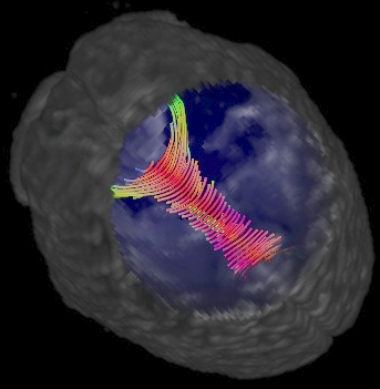
tomography (SPECT) and so on. In the case of epilepsy neurosurgery for
example, these images are used to identify brain regions to guide
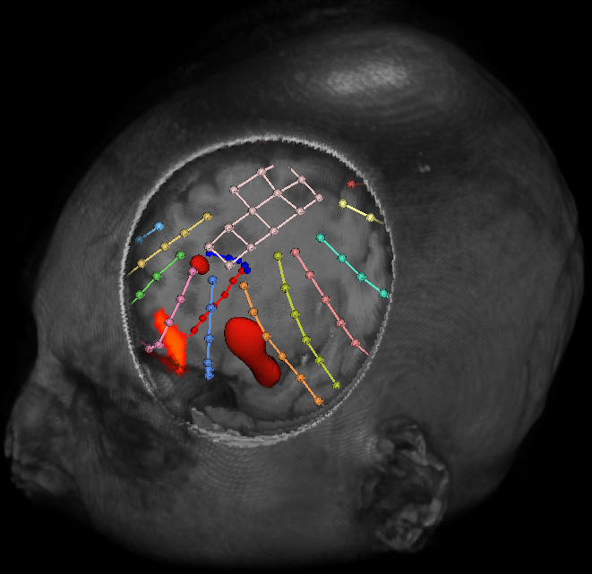
intracranial electrode implantation and resection. Generally, such data is
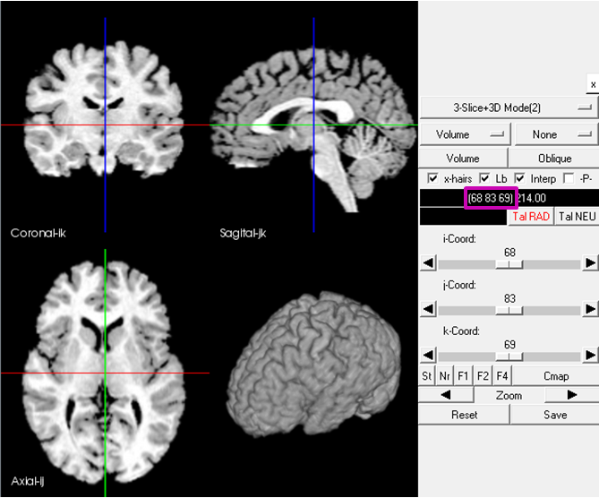
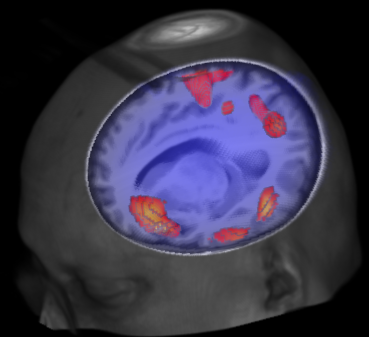
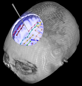
visualized using 2D slices and in some cases using a 3D volume rendering
along with the functional imaging results. Visualizing the activation
region effectively by still preserving sufficient surrounding brain regions
for context is exceedingly important to neurologists and surgeons.
We present novel interaction techniques for visualization of multimodal
data to facilitate improved exploration and planning for neurosurgery. We
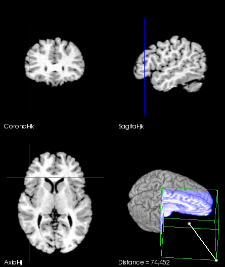
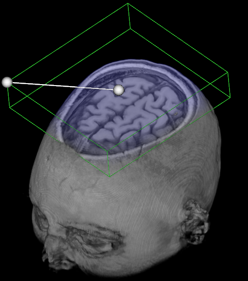
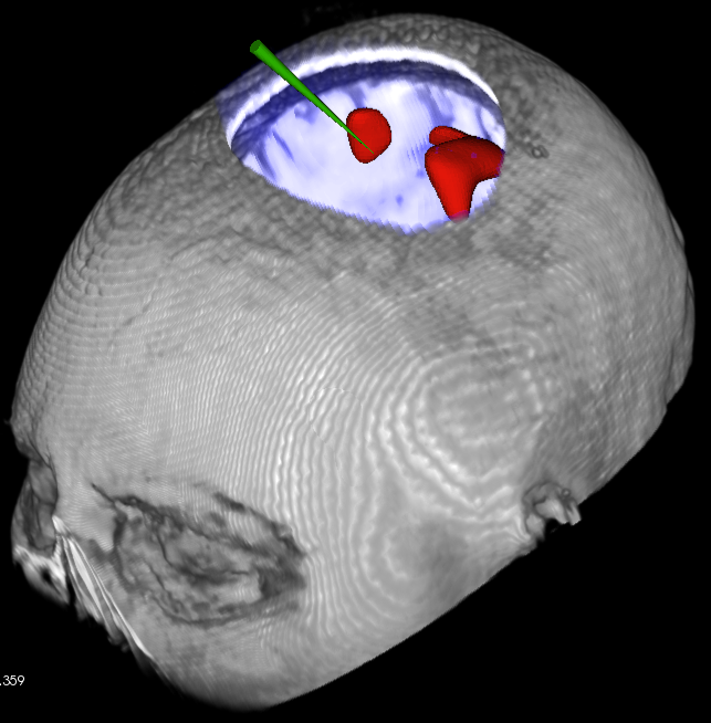
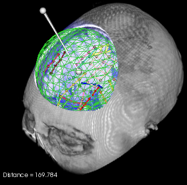
extended the line widget from VTK to allow surgeons to control the shape of
the region of the brain that they can visually crop away during exploration
and surgery. We allow simple spherical, cubical, ellipsoidal and
cylindrical (probe aligned cuts) for exploration purposes. In addition we
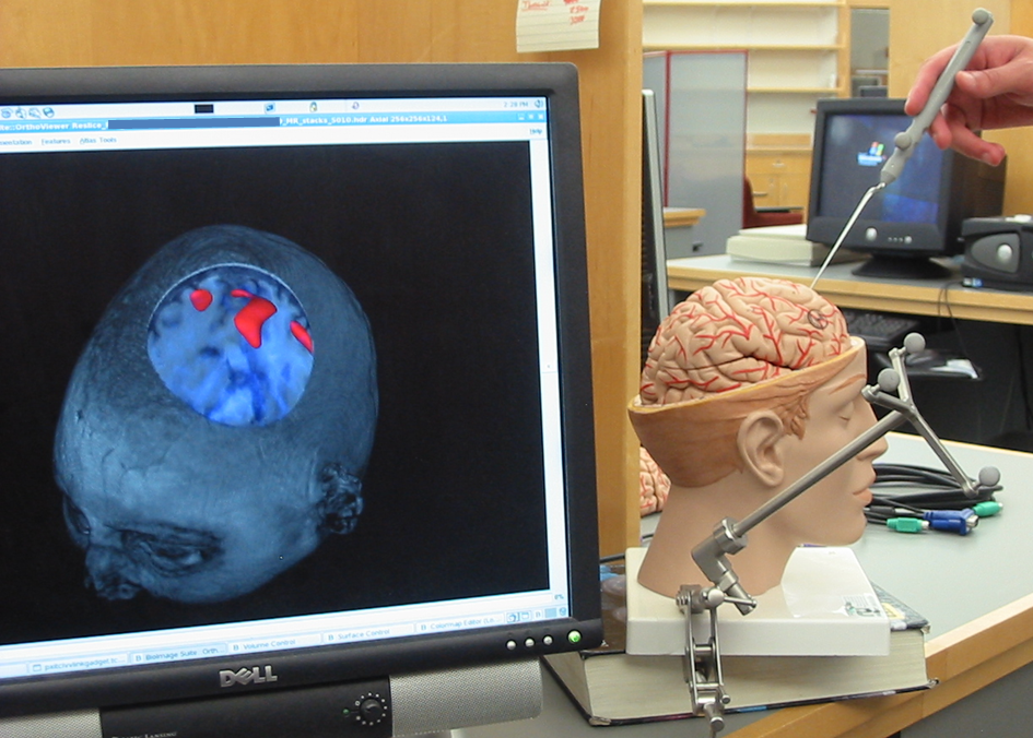
integrate the cropping tool with the image-guided navigation system used
for epilepsy neurosurgery. We are currently investigating the use of these
new tools in surgical planning and based on further feedback from our
neurosurgeons we will integrate them into the setup used for image-guided
neurosurgery.
 WMV(20M)